Separating chromosomes in fused cells
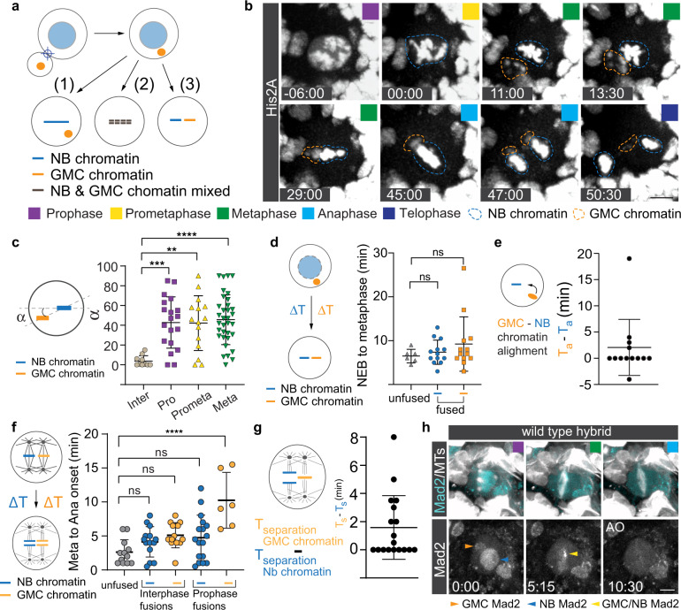
Figure 1 reproduced from Sunchu B., Lee N. M., Taylor J. A., Segura R. C., Roubinet C. & Cabernard C. (2022). Asymmetric chromatin retention and nuclear envelopes separate chromosomes in fused cells in vivo. Communications Biology 5, 953. doi:10.1038/s42003-022-03874-z. © 2022 The Author(s). Licensed under CC BY 4.0.
Every time a cell divides, it has to distribute a complete copy of its chromosomes to each daughter cell. This is normally straightforward because a cell only has one genome. However, after events such as cell fusion, two genetically distinct chromosome sets suddenly have to be managed within the same cell, and getting it wrong leads to chromosome missegregation and aneuploidy. Over a century ago, Aichel proposed that unregulated cell fusion could even seed tumors, which raises a simple question: how does the cell keep the two genomes apart?
To study this, we developed a hybrid-cell model in the Drosophila larval brain by briefly fusing a neural stem cell (neuroblast) with one of its differentiating daughters (a ganglion mother cell, GMC) using a focused 532 nm laser pulse. This created cells containing two distinct genomes, allowing us to watch how they behaved during mitosis.
The outcome depended on when the fusion happened. When cells were fused during interphase or early prophase, the neuroblast and GMC chromosomes stayed largely separated rather than mixing: each genome formed its own bipolar spindle before eventually aligning on a common metaphase plate, so the two chromosome sets could segregate independently within the same hybrid cell. Fusions induced later, in metaphase or anaphase, usually left the incoming GMC chromatin unable to align.
We found that this separation begins well before mitosis. During interphase, the neuroblast’s active apical microtubule-organising centre (MTOC) keeps its own centromeres positioned near the apical cortex, physically biasing them away from the incoming GMC chromosomes. Disrupting this asymmetry by depleting Centrobin caused the two centromere populations to come together much earlier than in control cells.
A second layer of separation comes from the nuclear envelope. Since neuroblasts undergo semi-closed mitosis, each genome initially remains enclosed within its own nuclear envelope, creating a physical barrier that keeps the chromosome sets apart until roughly metaphase.
Together, these findings show that hybrid cells use both asymmetric chromatin positioning during interphase and separate nuclear envelopes to keep distinct genomes apart before mitosis, with no evidence of active chromosome “recognition”. Segregation was still error-prone, producing lagging chromosomes, anaphase bridges and micronuclei, consistent with Aichel’s century-old idea that cell fusion can drive aneuploidy and tumor formation. The same mechanisms may also help preserve genomic organization at the first zygotic division.
As the paper’s second author, I laid much of the groundwork: through extensive troubleshooting I helped establish the laser-fusion system and uncovered much of the timing-dependent behavior at its heart, and I built the foundation for the processing and analysis of the imaging data behind these results.
To learn more: 10.1038/s42003-022-03874-z