Kalium channelrhodopsins
What KCRs are
Optogenetics lets us silence neurons with light, but the standard inhibitory tools, light-gated chloride channels (anion channelrhodopsins, ACRs), have an important limitation. In cells or subcellular compartments with high intracellular chloride, they can actually excite neurons instead of silencing them. Kalium channelrhodopsins (KCRs) avoid this by conducting potassium rather than chloride, hyperpolarizing neurons regardless of their chloride gradient.
This study evaluated KCRs as optogenetic silencers across Drosophila, C. elegans, and zebrafish, benchmarking them directly against the widely used chloride channel ACR1. An engineered KCR1 variant with improved membrane trafficking matched ACR1’s silencing performance while showing lower toxicity and better performance in putative high-chloride cells, establishing KCRs as a promising next-generation inhibitory tool.
The C. elegans arm
My contribution focused on the C. elegans behavioral experiments, where I tested whether KCRs could silence neurons by measuring changes in locomotion. Since there was no C. elegans behavioral lab locally and dedicated worm-tracking systems were prohibitively expensive, I built both the behavioral assay and tracking system from the ground up, starting with little more than a stereoscope and camera.
What was already out there
Before building anything, I surveyed the automated worm-tracking landscape. Our lab’s existing CRITTA system, built for insects, was unsuitable: the low-contrast stereoscope video gave too little foreground–background separation, and its single centroid-and-heading output cannot represent a body that constantly bends and coils. Among purpose-built worm trackers, classical computer-vision pose estimators need high spatial resolution2–8 and adequate contrast5,9–11 to extract limb edges reliably; low contrast defeats edge detectors regardless of pixel count8,12–15, and coiling or overlap forces most trackers to discard those events or require manual re-linking4. Skeleton-based systems such as Tierpsy Tracker16,17 extract more than 256 features but typically need ≥1280×1024 resolution with good contrast, and many high-end platforms require expensive specialized hardware, confocal microscopes, motorized stages, or high-speed cameras1,18–24. Deep-learning trackers are more robust to image-quality variation, including YOLO-based9,25 and Swin Transformer26 architectures. I tested classical tools (Tierpsy Tracker16,17 and wrMTrck27), but they failed to detect worms in our low-contrast video, and worm-specific deep-learning systems were hard to implement at our resolution. I therefore chose DeepLabCut (DLC)28, a general-purpose pose-estimation framework I could train on our own data. Full comparisons are in Tables D.4 and D.5 below.
Full comparison — Tables D.4 & D.5 (classical & machine-learning worm trackers)
Table D.4. Classical computer-vision methods for C. elegans tracking. Imaging requirements, detection and segmentation, skeleton extraction, tracking algorithm, and behavioral feature outputs. GUI, graphical user interface.
| Tracker | Required Resolution | Camera/Setup | Single/Multi Organism | GUI | Detection Method | Segmentation Method | Skeleton Extraction | Tracking Algorithm | Overlap | Features |
|---|---|---|---|---|---|---|---|---|---|---|
| Worm Tracker 2.0 29 | 640x480 | Motorized x-y stage, DinoLite AM413T camera with zoom magnification | Single | Yes | Thresholding + morphology | Morphological operations | 49-point skeleton | Stage tracking (follows worm) |
N/A (single worm) |
702 features |
| Nemo 30 | 800x600 | Zeiss Stemi SV11 stereomicroscope + Moticam 2000 CCD | Single | Yes | Thresholding | Morphological operations + skeletonization | Yes (7 segments) | Frame-to-frame linking |
N/A (single worm) |
Speed, direction change, wavelength, amplitude, body thickness |
| Parallel Worm Tracker 2 | 640x480 pixels | DCAM-compatible video camera (XCD-900, Sony), + zoom lens with C -mount adapter | Multiple (parallel tracking) | Yes | Thresholding | Thresholding | No (centroid only) | Multi-object linking | N/A | Speed, paralysis fraction |
| Track-A-Worm 4 | 1920x1080 | Stereomicroscope, motorized x-y stage, camera | Single | Yes | Thresholding + corner detection | Thresholding + corner detection | 13-point cubic spline | Stage tracking (re-centers at 1-sec intervals) |
N/A (single worm) |
Speed, distance, direction, bending frequency, amplitude |
| Multi-Worm Tracker 3 | 2,352 x 1,728 pixels | Dalsa Falcon 4M30, frame grabber, backlight | 5-120 | Lab View | Thresholding + flood-fill | Threshold + flood-fill from dark pixels | 11-point spine | Real-time object association | Partial (loses identity upon collision) | Speed, angular speed, area, spine length, curvature, body bends |
| CeleST 31 | 0.02 mm/pixel minimum, 696 x 520pixel | Digital Camera | 1-5 | Yes | Thresholding + curvature | Curvature-based centerline | Yes | Per-frame | Partial | 10 swimming parameters |
| wrMTrck 27 | 640x480 (flexible) | USB microscope | Multiple (~8) | Yes | Max entropy thresholding | Thresholding | No (ellipse fitting) | Centroid linking | Breaks tracks | Size, velocity, BLPS, BBPS |
| CoLBeRT 32 | ~30 µm (swimming), ~5 µm limit (crawling) | 10X Nikon Eclipse TE2000-U with PhotonFocus MV2-D1280-640CL (high-speed) | Single | Yes | Thresholding + boundary detection | Thresholding (filtered image) | Yes - centerline with 100 segments | MindControl: Stage-tracking + boundary/centerline extraction | N/A | Centroid, curvature, bending wave speed, head/tail position |
| WF-NTP 5 | ≥6 megapixel recommended | 6MP camera (Edmund Optics, model no. GS3-U3-41C6M-C) + flat-field illumination | Up to 5,000 | Yes | Adaptive Gaussian thresholding | Gaussian thresholding + morphology | Skeletonization (for centroid) | Collision-aware tracking | Partial | Speed, paralysis, area |
| 3D-worm tracker 33 | 550x550 (trajectory analysis); 1024x1024 + 1600x1200 (kinematic) | FASTCAM SA1.1 + PCO.1600 dual cameras (stereoscopic) | Single | No | Thresholding + stereomatching | Thresholding + stereo reconstruction | Yes (3D skeleton, 13 sections) | Manual stage control + per-frame | Detects Coiling | 3D trajectory, 3D posture, bending vectors |
| Tierpsy Tracker 16,17 | >75pixels/mm or 1280 x 1024 | Standard camera + light source | Single and multi-worm | Yes | Adaptive thresholding | Adaptive thresholding + size filtering | segWorm algorithm (from Worm Tracker 2.0) - identifies contour points with highest curvature, divides contour into ventral/dorsal, calculates midline | Frame-to-frame trajectory joining | No | 256+ features |
Table D.5. Machine-learning methods for C. elegans tracking. Deep-learning approaches for worms and other organisms: architectures for detection, segmentation, and pose estimation; worm-specific tools and adaptable general-purpose frameworks. CNN, convolutional neural network; YOLO, You Only Look Once; SORT, Simple Online and Realtime Tracking; ReID, re-identification.
| Tracker | Required Resolution | Camera/Setup | Single/Multi Organism | GUI | Detection Method | Segmentation Method | Skeleton Extraction | Tracking Algorithm | Overlap | Features |
|---|---|---|---|---|---|---|---|---|---|---|
|
Deep Tangle7 |
512 x 512 resolution | Standard microscopy | Multiple, up to 6000 | No | Single-stage detection model with ResNet backbone | None - bypasses and uses neural networks | Direct prediction by CNN | Linear assignment problem with directed metric |
Limited coiling |
centerline angle, center-of-mass speed, curvature |
|
Deep Tangle Crawl 10 |
12.4 µm/pixel | Megapixel camera arrays | Multiple | Yes | DeepTangle (modified ResNet backbone + YOLO-style grid output) | None - bypasses pixel segmentation entirely | Direct CNN prediction | Built-in temporal tracking (11-frame context) | Yes | 256 features similar to Tierpsy |
| Deep-Worm-Tracker 9 | 0.8 MP, 2 MP, 5 MP | CMOS camera + Leica stereo microscope | Multiple | No | YOLOv5 object detection | Threshold-based | Yes - morphological skeletonization | Strong SORT (appearance + motion + Kalman + Hungarian Assignment) | Partial | Trajectory |
|
Worm Swin 26 |
800px x 1333px | N/A | Multiple | No | Hybrid Task Cascade (HTC) + Swin Transformer backbone | Instance segmentation (end-to-end deep learning) | No -outputs masks only | Simple IoU-based matching: Match highest overlapping masks between consecutive frames | Yes | Bounding boxes + instance segmentation masks |
|
Worm YOLO 25 |
640 x 480 | Standard microscopy | Multiple | No | YOLO + RepLKNet | Instance segmentation (end-to-end deep learning) | Yes - post-processing algorithm on segmented masks | BoT-SORT + Kalman filter + Hungarian algorithm + ReID appearance features | Partial | Head/body/tail bend count, max bend speed, body length, frames where head-tail distance < 20% body length |
| YOLOv8 34 | 1024 x 1024 pixels | Olympus SZX7 stereo microscope + DP20 camera; Custom microscope + Mshiwi SUA134GC/M industrial camera | Multiple | N/A | CSPDarknet backbone + PANet neck + Detection head | YOLO-style bounding box detection | None - indirect via bounding box | ByteTrack + Kalman filter | Partial | ~7 types |
|
Worm Pose 35 |
Not specified | Standard microscopy setup | Single | No | CNN-based | Generative model + CNN | Yes (pose estimation, reverse skeletonization) | Per-frame pose estimation |
Detects Coiling (single worm) |
Posture angles |
| Deep Lab Cut (DLC) 28 | minimum 640 x 480 (low-res) | N/A | Multiple | Yes | DeeperCut feature detectors (ResNet-50 + deconvolutional layers) | Direct keypoint detection | User-defined keypoints - not automatic skeleton | Frame-by-frame detection | N/A | x, y coords + likelihood per bodypart per frame |
|
Social LEAP Estimates Animal Poses (SLEAP) 36 |
1,024 x 1,024 pixels | No specific camera required | Multiple | Yes | Deep learning-based pose estimation using CNN | Confidence maps (2D Gaussians); multi-class segmentation maps for ID models | User-defined nodes and edges; skeleton modeled as directed tree | Flow-shift or appearance-based ID classification with optimal assignment | Yes | x, y coords + point/instance/tracking scores per node per track per frame |
So we built our own
As none of the available systems worked for a low-contrast stereoscope setup, I designed and built a dedicated C. elegans optogenetics and tracking rig. It combined custom CNC-milled chambers, magnetic modular components, a 3D-printed camera adapter, infrared and optogenetic illumination, and a DeepLabCut tracker trained on our own recordings.
Click here to see how we made it → the C. elegans tracking rig
What we found
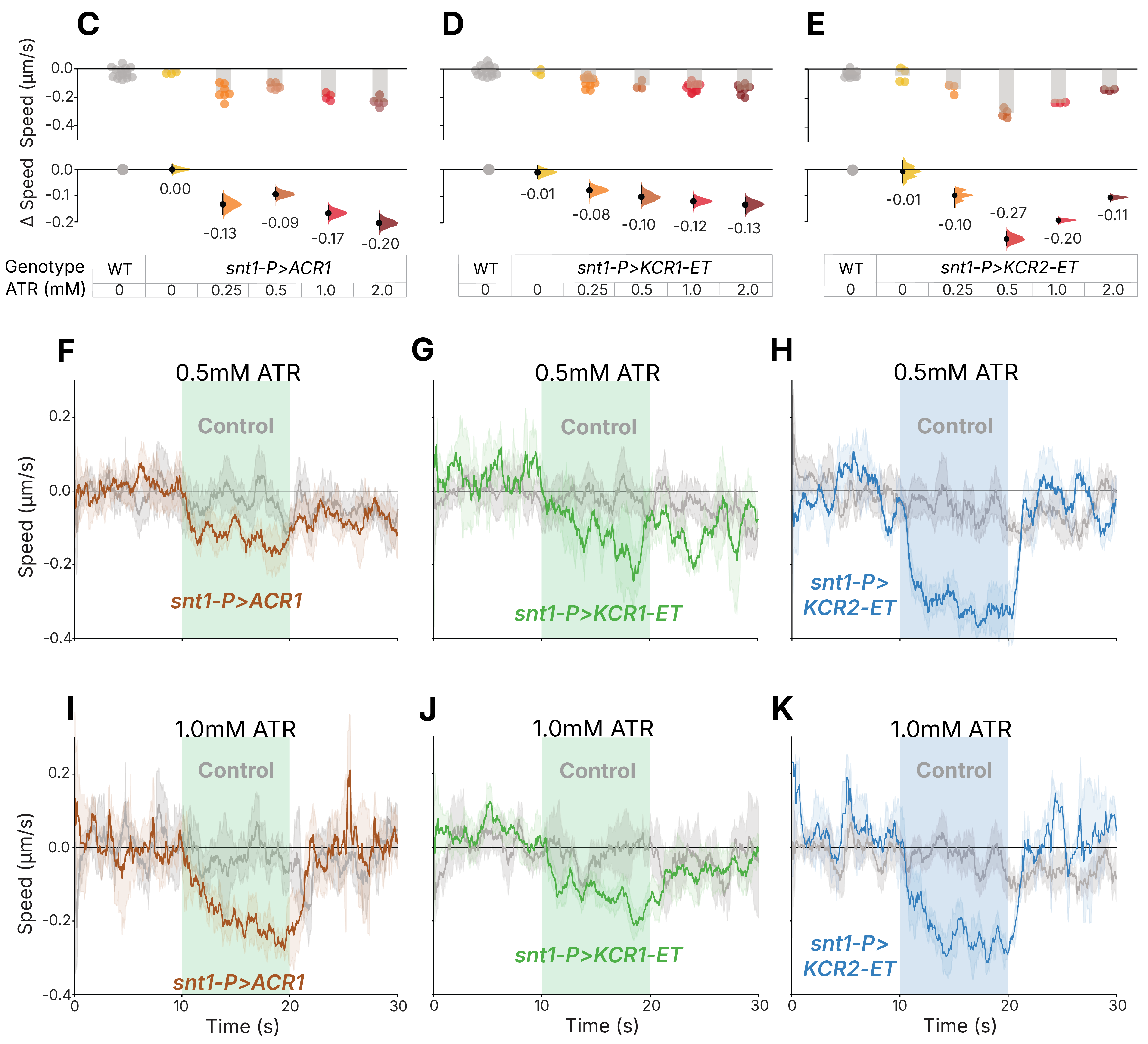
A crop of the paper’s Figure 5 showing the C. elegans results — the part I worked on: pan-neuronally expressed opsins reduce worm movement during green illumination, with rapid recovery once the light is off. Figure 5 (cropped to the C. elegans results) from Ott S., Xu S., Lee N., Hong I., Anns J., Suresh D. D., Zhang Z., Zhang X., Harion R., Ye W., Chandramouli V., Jesuthasan S., Saheki Y. & Claridge-Chang A. (2024). Kalium channelrhodopsins effectively inhibit neurons. Nature Communications 15, 3480. doi:10.1038/s41467-024-47203-w. © 2024 The Author(s). Licensed under CC BY 4.0.
To measure neuronal silencing, worms expressing each opsin pan-neuronally were reared on different concentrations of all-trans-retinal (ATR) and recorded on the custom rig. Every opsin line slowed markedly during green-light stimulation, while the enhanced-trafficking (ET) tag improved membrane targeting in worms just as it had in flies and cultured cells. Overall, KCR2-ET performed best, followed by ACR1 and KCR1-ET, although the ranking depended on ATR concentration. All animals recovered rapidly once the light was turned off, with no meaningful differences in recovery time between genotypes.
Across all three model organisms, the trafficking-enhanced KCR1 matched ACR1’s silencing performance while causing less developmental toxicity and performing better in putative high-chloride cells, supporting KCRs as a next-generation optogenetic silencer.
The work was covered in The Straits Times (2024).
To learn more: 10.1038/s41467-024-47203-w
References
1. Stephens, G. J., Johnson-Kerner, B., Bialek, W. & Ryu, W. S. Dimensionality and dynamics in the behavior of C. elegans. PLoS Comput. Biol. 4, e1000028 (2008).
2. Ramot, D., Johnson, B. E., Berry, T. L., Jr, Carnell, L. & Goodman, M. B. The Parallel Worm Tracker: a platform for measuring average speed and drug-induced paralysis in nematodes. PLoS One 3, e2208 (2008).
3. Swierczek, N. A., Giles, A. C., Rankin, C. H. & Kerr, R. A. High-throughput behavioral analysis in C. elegans. Nat. Methods 8, 592–598 (2011).
4. Wang, S. J. & Wang, Z.-W. Track-a-worm, an open-source system for quantitative assessment of C. elegans locomotory and bending behavior. PLoS One 8, e69653 (2013).
5. Koopman, M. et al. Assessing motor-related phenotypes of Caenorhabditis elegans with the wide field-of-view nematode tracking platform. Nat. Protoc. 15, 2071–2106 (2020).
6. Layana Castro, P. E., Puchalt, J. C., García Garví, A. & Sánchez-Salmerón, A.-J. Caenorhabditis elegans multi-tracker based on a modified skeleton algorithm. Sensors (Basel) 21, 5622 (2021).
7. Alonso, A. & Kirkegaard, J. B. Fast detection of slender bodies in high density microscopy data. Commun. Biol. 6, 754 (2023).
8. Layana Castro, P. E., García Garví, A., Navarro Moya, F. & Sánchez-Salmerón, A.-J. Skeletonizing Caenorhabditis elegans based on U-Net architectures trained with a multi-worm low-resolution synthetic dataset. Int. J. Comput. Vis. 131, 2408–2424 (2023).
9. Banerjee, S. C., Khan, K. A. & Sharma, R. Deep-worm-tracker: Deep learning methods for accurate detection and tracking for behavioral studies in C. elegans. Appl. Anim. Behav. Sci. 266, 106024 (2023).
10. Weheliye, W. H. et al. A neural network model enables worm tracking in challenging conditions and increases signal-to-noise ratio in phenotypic screens. PLoS Comput. Biol. 21, e1013345 (2025).
11. Benji, N. A guide to robust edge detection with OpenCV. Medium https://medium.com/@noel.benji/a-guide-to-robust-edge-detection-with-opencv-1d703506e014 (2025).
12. Roussel, N., Sprenger, J., Tappan, S. J. & Glaser, J. R. Robust tracking and quantification of C. elegans body shape and locomotion through coiling, entanglement, and omega bends. Worm 3, e982437 (2014).
13. Spontón, H. & Cardelino, J. A review of classic edge detectors. Image Process. Line 5, 90–123 (2015).
14. Ofir, N. et al. On detection of faint edges in noisy images. arXiv [cs.CV] (2017).
15. Muntarina, K., Mostafiz, R., Shorif, S. B. & Shorif Uddin, M. Deep learning-based edge detection for random natural images. Neurosci. Inform. 5, 100183 (2025).
16. Javer, A. et al. An open-source platform for analyzing and sharing worm-behavior data. Nat. Methods 15, 645–646 (2018).
17. Barlow, I. L. et al. Megapixel camera arrays enable high-resolution animal tracking in multiwell plates. Commun. Biol. 5, 253 (2022).
18. Wang, W., Sun, Y., Dixon, S. J., Alexander, M. & Roy, P. J. An automated micropositioning system for investigating C. elegans locomotive behavior. J. Lab. Autom. 14, 269–276 (2009).
19. Stirman, J. N., Brauner, M., Gottschalk, A. & Lu, H. High-throughput study of synaptic transmission at the neuromuscular junction enabled by optogenetics and microfluidics. J. Neurosci. Methods 191, 90–93 (2010).
20. Stirman, J. N. et al. Real-time multimodal optical control of neurons and muscles in freely behaving Caenorhabditis elegans. Nat. Methods 8, 153–158 (2011).
21. Leifer, A. M., Fang-Yen, C., Gershow, M., Alkema, M. J. & Samuel, A. D. T. Optogenetic manipulation of neural activity in freely moving Caenorhabditis elegans. Nat. Methods 8, 147–152 (2011).
22. Faumont, S. et al. An image-free opto-mechanical system for creating virtual environments and imaging neuronal activity in freely moving Caenorhabditis elegans. PLoS One 6, e24666 (2011).
23. Gengyo-Ando, K. et al. A new platform for long-term tracking and recording of neural activity and simultaneous optogenetic control in freely behaving Caenorhabditis elegans. J. Neurosci. Methods 286, 56–68 (2017).
24. Qiu, Z. et al. An integrated platform enabling optogenetic illumination of Caenorhabditis elegans neurons and muscular force measurement in microstructured environments. Biomicrofluidics 9, 014123 (2015).
25. Dong, B. & Chen, W. A high precision method of segmenting complex postures in Caenorhabditis elegans and deep phenotyping to analyze lifespan. Sci. Rep. 15, 8870 (2025).
26. Deserno, M. & Bozek, K. WormSwin: Instance segmentation of C. elegans using vision transformer. Sci. Rep. 13, 11021 (2023).
27. Nussbaum-Krammer, C. I., Neto, M. F., Brielmann, R. M., Pedersen, J. S. & Morimoto, R. I. Investigating the spreading and toxicity of prion-like proteins using the metazoan model organism C. elegans. J. Vis. Exp. 52321 (2015).
28. Mathis, A. et al. DeepLabCut: markerless pose estimation of user-defined body parts with deep learning. Nat. Neurosci. 21, 1281–1289 (2018).
29. Yemini, E., Jucikas, T., Grundy, L. J., Brown, A. E. X. & Schafer, W. R. A database of Caenorhabditis elegans behavioral phenotypes. Nat. Methods 10, 877–879 (2013).
30. Tsibidis, G. D. & Tavernarakis, N. Nemo: a computational tool for analyzing nematode locomotion. BMC Neurosci. 8, 86 (2007).
31. Restif, C. et al. CeleST: computer vision software for quantitative analysis of C. elegans swim behavior reveals novel features of locomotion. PLoS Comput. Biol. 10, e1003702 (2014).
32. Yu, X. et al. Fast deep neural correspondence for tracking and identifying neurons in C. elegans using semi-synthetic training. Elife 10, e66410 (2021).
33. Kwon, N., Pyo, J., Lee, S.-J. & Je, J. H. 3-D worm tracker for freely moving C. elegans. PLoS One 8, e57484 (2013).
34. Liu, X. et al. Automated C. elegans behavior analysis via deep learning-based detection and tracking. PLoS Comput. Biol. 21, e1013707 (2025).
35. Hebert, L., Ahamed, T., Costa, A. C., O’Shaughnessy, L. & Stephens, G. J. WormPose: Image synthesis and convolutional networks for pose estimation in C. elegans. PLoS Comput. Biol. 17, e1008914 (2021).
36. Pereira, T. D. et al. SLEAP: A deep learning system for multi-animal pose tracking. Nat. Methods 19, 486–495 (2022).