Sibling cell size asymmetry
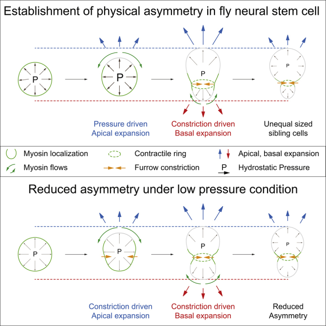
Graphical abstract reproduced from Pham T.T., Monnard A., Helenius J., Lund E., Lee N., Müller D.J. & Cabernard C. (2019). Spatiotemporally Controlled Myosin Relocalization and Internal Pressure Generate Sibling Cell Size Asymmetry. iScience 13, 9–19. doi:10.1016/j.isci.2019.02.002. © 2019 The Author(s). Reproduced under CC BY-NC-ND 4.0.
Many stem cells divide asymmetrically, producing two daughters that are different not only in fate but also in size. In Drosophila neural stem cells, the larger daughter remains a stem cell while the smaller daughter begins differentiating. How cells reliably generate this size difference during every division has remained an open question.
Using atomic force microscopy alongside live imaging of dividing Drosophila neural stem cells, we measured the physical forces at work. Two of them drive the unequal division: intracellular hydrostatic pressure and cortical actomyosin contractility, each acting at a different moment and a different part of the cell.
The timing is what makes it work. Just after anaphase onset, hydrostatic pressure peaks and drives expansion of the apical cortex, the future neuroblast. A little later, contractile tension rises at the cleavage furrow and myosin relocalizes, driving basal expansion. Because the two forces act on different timescales and different sides of the cell, the division ends with a larger self-renewing neuroblast and a smaller differentiating GMC.
The system is also surprisingly robust. When we perturbed one of these inputs, the cell did not simply lose its asymmetry: the cleavage furrow repositioned to compensate, shifting so that the two daughters still ended up different sizes. That redundancy is what makes the size difference so reliable from one division to the next.
Our findings show that asymmetric cell division emerges from the combined action of intracellular pressure and cortical mechanics, providing a physical explanation for how neural stem cells reliably generate daughters of different sizes.
This was an early collaboration from my developmental biology years, and as a co-author it was a formative lesson for me in pairing quantitative biophysics with live imaging to dissect a mechanism neither approach could resolve on its own.
To learn more: 10.1016/j.isci.2019.02.002